Enlarged Blind Spot Syndrome
Enlarged blind spot syndrome. Ophthalmoscopic features included uveitis mild optic nerve swelling granularity of macular pigment subretinal white dots and peripapillary pigment disturbances. Unilateral enlargement of the blind spot. Dyschromatopsia and afferent pupil defects were prevalent.
This causes displacement of the peripapillary retina and thus an enlarged blind spot. This case was unique in that retinal findings consistent with MEWDS were present in addition to enlargement of the blind spot. The related entity idiopathic enlargement of the blind spot syndrome shares demographic features with MEWDS with the noted absence of retinal lesions.
Unilateral blind spot enlargement occurs as an isolated entity acute idiopathic blind spot enlargement or in association with other conditions such as multiple evanescent white dot syndrome multifocal choroiditis with panuveitis or punctate inner choroidopathy. It remains controversial whether blind spot enlargement. A review of 27 new cases.
The other symptoms and clinical presentation in this patient is more typical for acute idiopathic blind spot enlargement AIBSE syndrome or acute zonal occult outer retinopathy AZOOR. This led us to perform a MERG which demonstrated depression of retinal function OS and confirmed the diagnosis of AIBSEAZOOR. Patients with an enlarged blind spot typically have disc swelling.
Although the patient usually becomes asymptomatic the blind spot is slightly and permanently enlarged and. In the absence of marked disc swelling patients need a careful retinal examination to look for chorioretinitis. The disease affects the peripapillary retina and may cause an afferent pupillary defect.
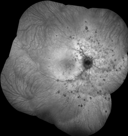
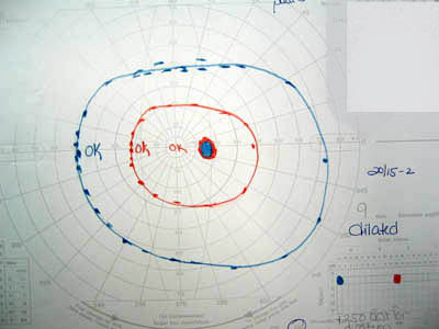
A The right fundus of a 16-year-old girl who was seen on April 13 1995 with a temporal scotoma and 2060 vision of 7 days duration. All patients had enlarged blind spots of variable size and density. There is disc congestion peripapillary outer retinal opacification small foveal deposits and B peripheral retinal spots characteristic of MEWDS.
1 In 1988 Fletcher et al 2 described a clinically distinct syndrome of blind spot enlargement in 7 patients who acutely developed photopsias and enlargement of the blind spot without optic disc swelling or choroiditis. If an eye with an enlarged blind spot is examined within 2 weeks of onset signs of a chorioretinal disease will usually be present.
A review of 27 new cases.
This causes displacement of the peripapillary retina and thus an enlarged blind spot. This case was unique in that retinal findings consistent with MEWDS were present in addition to enlargement of the blind spot. In patients with inflammatory disease of the retina and choroid blind spot enlargement corresponds to areas of retina and choroid that appear abnormal ophthalmoscopically. The related entity idiopathic enlargement of the blind spot syndrome shares demographic features with MEWDS with the noted absence of retinal lesions. Although the patient usually becomes asymptomatic the blind spot is slightly and permanently enlarged and. 1 In 1988 Fletcher et al 2 described a clinically distinct syndrome of blind spot enlargement in 7 patients who acutely developed photopsias and enlargement of the blind spot without optic disc swelling or choroiditis. It remains controversial whether blind spot enlargement. All patients had enlarged blind spots of variable size and density. The disease affects the peripapillary retina and may cause an afferent pupillary defect.
All patients had enlarged blind spots of variable size and density. In the absence of marked disc swelling patients need a careful retinal examination to look for chorioretinitis. Patients with an enlarged blind spot typically have disc swelling. Ophthalmoscopic features included uveitis mild optic nerve swelling granularity of macular pigment subretinal white dots and peripapillary pigment disturbances. The white dot syndromes are a group of inflammatory chorioretinopathies of unknown etiology which have in common a unique and characteristic appearance of multiple yellow-white lesions affecting multiple layers of the retina retinal pigment epithelium RPE choriocapillaris and the choroid. The disease affects the peripapillary retina and may cause an afferent pupillary defect. The other symptoms and clinical presentation in this patient is more typical for acute idiopathic blind spot enlargement AIBSE syndrome or acute zonal occult outer retinopathy AZOOR.


Post a Comment for "Enlarged Blind Spot Syndrome"